- Research,
- Future Health,
MRI quantif: The MRI of the future
Published on March 9, 2016 – Updated on February 28, 2024
The ‘MRI quantif’ project brings together researchers from Centrale Nantes - David Le Touzé (fluid mechanics, H2I research group of the LHEEA laboratory), Jérôme Idier and Saïd Moussaoui (signal processing, ADTSI research group of the IRCCyN laboratory) – and researchers from the Institut du Thorax, Nantes University Hospital - Jean-Michel Serfaty (radiology, MRI), Perrine Paul-Gilloteaux (signal processing) and Hubert Desal (radiology, MRI).

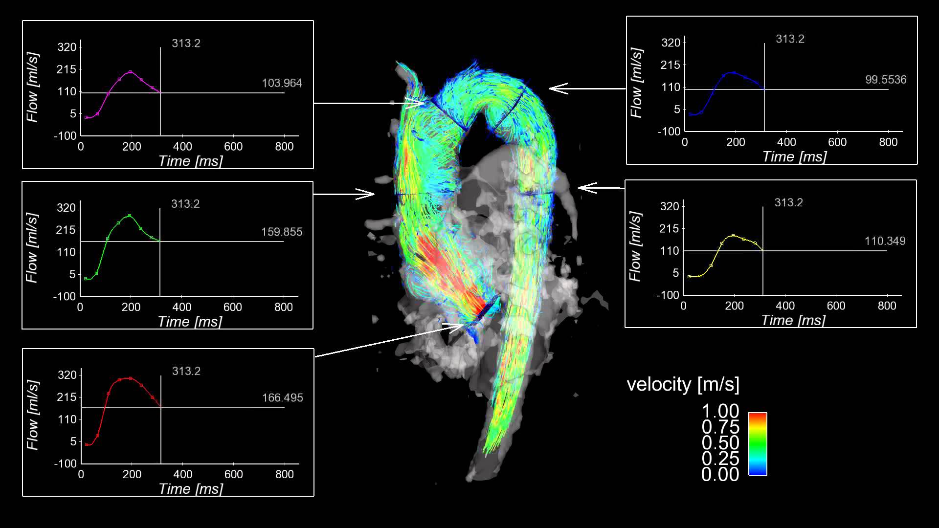
Instead of slice-by-slice images, today's 4D MRIs provide real-time volume visualisation, enabling blood flow visualisation throughout the part of the body under observation. Specialised procedures need to be developed in order to optimise these new MRIs, recently installed in Nantes University Hospital.
The researchers at Centrale Nantes are working on image and signal processing and applying their expertise in fluid mechanics to improve image resolution and accuracy. This expertise in fluid mechanics ensures that the processed MRI images produce blood flow visualisation of the vessel in question in agreement with the laws of physics. The first step is to validate this technique by artificially reproducing flows to be measured accurately by MRI and other reference techniques in parallel. The next step is to extract biomarkers: highly relevant data for the doctors. Thus, for example, using the new MRI machine to determine the geometry of the aorta and the blood flow within makes it possible to extract a biomarker, e.g. the friction produced by the blood against the vessel walls. This biomarker then helps the doctors to determine whether a patient suffers from a particular condition, and how advanced the condition is. Current research is focused on vessels such as the aorta, but will also ultimately address neurology.
Developing new processing methods for MRI data is already a major line of research at Centrale Nantes whose researchers are keen to enhance their research activities in the health sector. This ambitious, innovative and transdisciplinary project will be led by Centrale Nantes from 2016 to 2020 with the benefit of funding from the Pays de la Loire region.
In addition...
Centrale Nantes researcher, David Le Touzé, is working on other project leads at the same time.
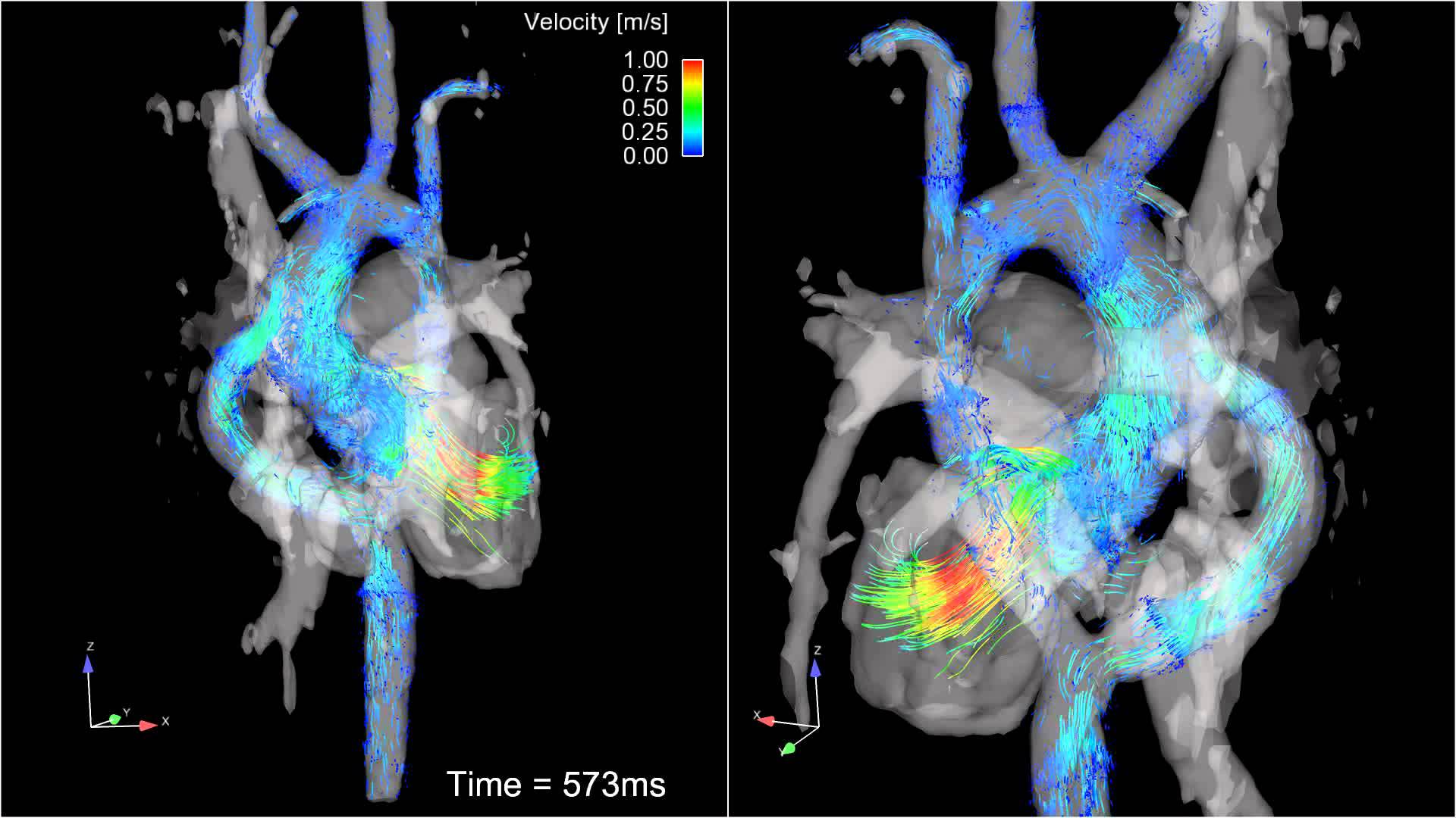
He is working in particular with the haemodynamics and cardiovascular centre of Nantes University Hospital, more specifically with Jean-Michel Serfaty's colleagues on blood flow in the heart and in blood vessels. The objective is to provide doctors with the benefit of their expertise and understanding of the behaviour of fluids to inform surgical procedures. Inserting springs into damaged vessels to keep them open or stapling heart valves to repair them ... these kinds of surgical procedure can alter the blood flow without doctors being fully aware of all the consequences. This is how the patient can benefit from the skills combination.
Another lead - this time in nuclear imaging. Work has started with the Cancer Institute of Nantes University Hospital in association with Jérôme Idier of the IRCCyN Laboratory. Their attention is focused on markers in sugar molecules, which are present in large quantities in cancer cells. The objective is to reduce the amount of radiotracers (short-lived and expensive) whilst improving image quality. A Centrale Nantes student is undertaking a 6-month final year internship on this subject.
Images from: Northwestern Radiology